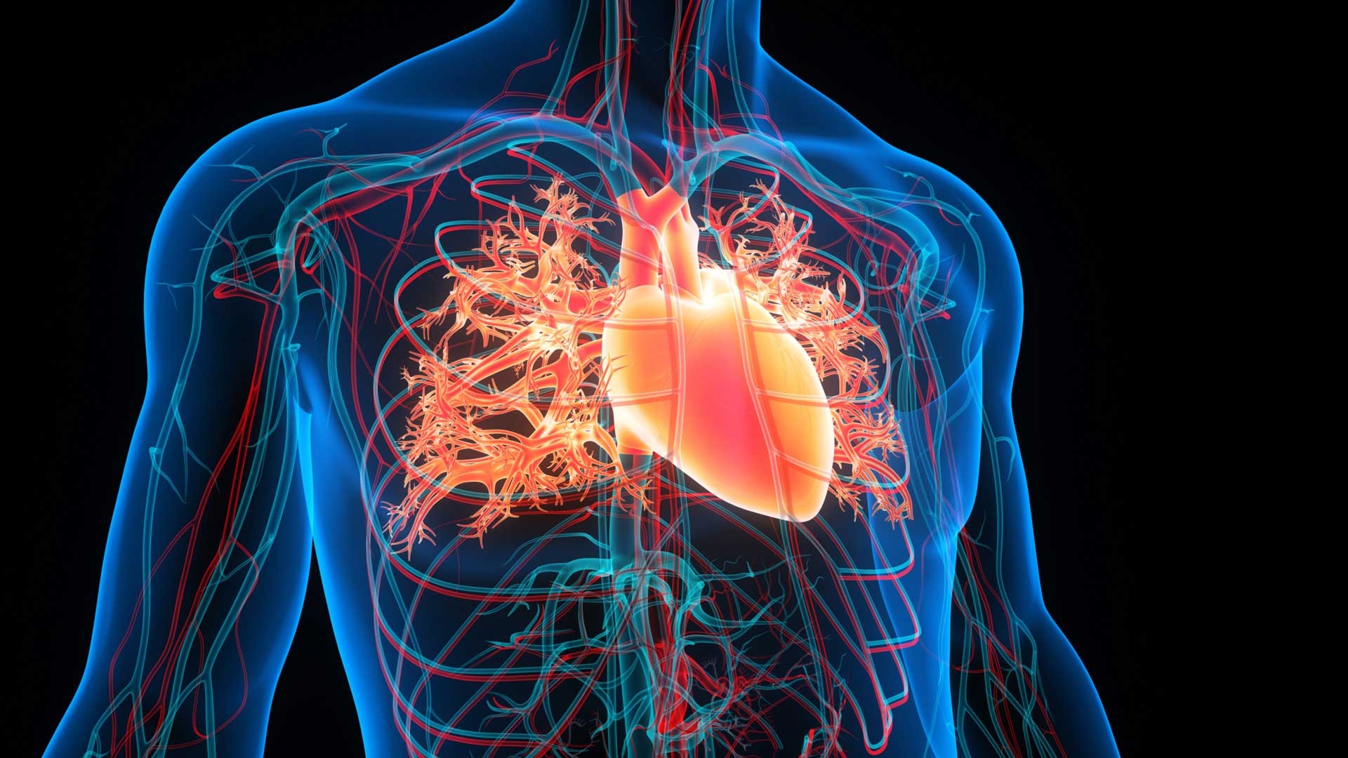
In the realm of modern diagnostics, few procedures offer as comprehensive a view as whole-body angiography. This imaging technique captures the intricate network of blood vessels that run throughout the body, offering doctors a full vascular map. Far beyond traditional scans that isolate single organs or regions, whole-body angiography presents a systemic perspective. It plays a crucial role in diagnosing, monitoring, and even preventing severe vascular events by uncovering underlying blood flow disturbances before they escalate into emergencies.
The Clinical Value Behind the Scan
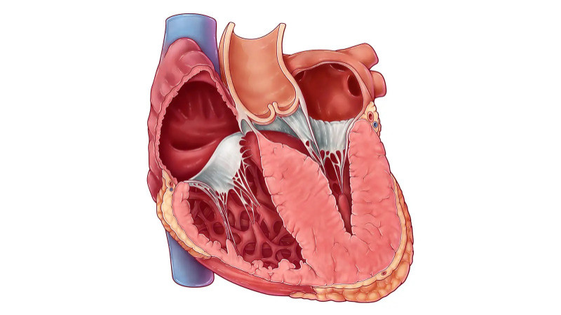
Whole-body angiography is typically employed when a patient presents with symptoms that suggest systemic vascular compromise. Conditions such as atherosclerosis, aneurysms, vascular malformations, or thromboembolic events often require such detailed imaging. Individuals with chronic diseases like diabetes, hypertension, or connective tissue disorders may also be candidates, given their elevated risk for vessel damage. The scan’s ability to detect blockages, narrowing, or abnormal vessel branching in multiple regions makes it a preferred tool for early diagnosis and holistic vascular assessment.
Preparing for the Imaging Process
Ahead of the scan, patients are usually advised to fast for several hours, especially when contrast dye is involved. This precaution helps minimize any gastrointestinal side effects and ensures clearer imaging results. Blood tests are often ordered beforehand to evaluate kidney function, as the dye used in CT angiography can strain renal systems in susceptible individuals. Those undergoing MRI angiography are screened for metallic implants or devices, since the strong magnetic fields may interfere with safety or image quality.
A simple hospital gown replaces everyday clothing to avoid any metal artifacts. Medical history, allergies—particularly to iodine or gadolinium—and any history of contrast reactions are reviewed thoroughly before the procedure begins. These steps ensure that the process proceeds safely and efficiently.
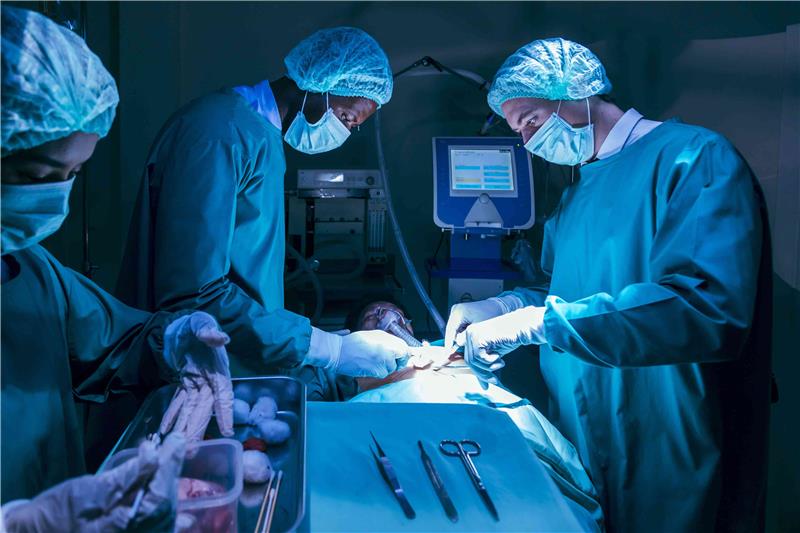
Inside the Scan Room
The angiography procedure begins with the insertion of an intravenous line, typically into a vein in the arm. Through this line, a contrast agent is introduced to enhance the visibility of blood vessels during imaging. Once administered, the contrast travels through the bloodstream, making arteries and veins more distinguishable.
The patient is then positioned on a motorized table that slides slowly into a scanner—either a CT or MRI machine. CT-based angiography uses X-ray technology to create cross-sectional images, while MRI angiography relies on powerful magnetic fields. The scan is painless and non-invasive, though it may require the patient to remain still or hold their breath momentarily to capture precise images. The entire process generally takes 30 to 60 minutes, depending on the scanner type and image resolution required.
Some individuals notice a temporary sensation of warmth or a metallic taste when the contrast is administered. These sensations are normal and subside quickly. Throughout the procedure, trained technicians monitor from an adjacent control room, maintaining constant communication for reassurance and guidance
What Happens After the Scan
Following the scan, a short observation period is standard, especially if a contrast agent was used. This allows the medical team to ensure there are no immediate allergic reactions or complications. Most patients are encouraged to drink plenty of water afterward, aiding the kidneys in flushing out the contrast material naturally.
There are no lasting physical effects from the scan itself, and most individuals can resume their regular activities within the same day. In rare cases, mild dizziness or nausea may occur, usually resolving without any intervention
Interpreting the Images
Once the imaging is complete, the digital data is sent to a radiologist with expertise in vascular anatomy. These specialists analyze the scan for any signs of blockages, vessel narrowing, aneurysms, or vascular anomalies. The report is then forwarded to the referring physician, who correlates the imaging findings with clinical symptoms to determine the next steps.
Whether the outcome calls for medication, lifestyle changes, further diagnostic tests, or surgical intervention, this imaging technique provides the clarity needed for precise decision-making
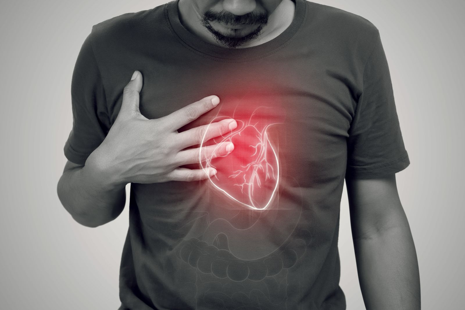
Prevention Through Precision
One of the most significant contributions of whole-body angiography lies in its ability to prevent adverse events. Many vascular conditions develop quietly and progress without symptoms until they reach a critical stage. Detecting these conditions early can lead to interventions that avert heart attacks, strokes, or organ failure. For high-risk individuals, especially those with a strong family history of cardiovascular disease, this scan offers not just diagnosis—but a preventative edge.
Balancing Diagnostic Benefits with Safety
As with any diagnostic tool, whole-body angiography carries minor risks. The use of contrast dye can occasionally trigger allergic responses or impact kidney function, although these risks are carefully screened in advance. CT angiography involves radiation exposure, but current machines use optimized low-dose protocols. MRI angiography, on the other hand, avoids radiation entirely and is considered safer for younger individuals or those requiring repeated scans.
The medical team assesses whether the benefits of the scan outweigh potential risks, ensuring that the procedure is medically justified and tailored to each patient’s condition.
Conclusion:
Whole-body angiography provides more than just images; it offers a detailed blueprint of the body's vascular infrastructure. Its ability to uncover early signs of vascular compromise, map disease progression, and guide targeted interventions makes it an invaluable tool in modern medicine. For clinicians, it opens a clear path to accurate diagnosis and treatment. For patients, it brings clarity, confidence, and an opportunity to safeguard future health.